3D Mammogram

Contact Us

812-376-5064

2326 18th Street
Columbus, IN

Get Directions Schedule A Mammogram Contact Us Online

Digital Breast Tomosynthesis (tomo)

 A 3D mammogram (also known as digital breast tomosynthesis or "tomo") is an exam that allows doctors to see masses and distortions associated with cancers significantly more clearly than conventional 2D mammograms. Instead of viewing all of the complexities of your breast tissue in a flat image, as with conventional mammography, fine details are more visible and no longer hidden by the tissue above or below.

What is a 3D mammogram?

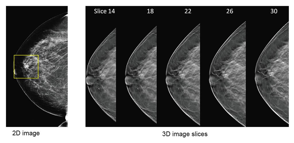
This exam, also known as digital breast tomosynthesis or "tomo," is an advanced technology in the fight against breast cancer that allows doctors to examine your breast tissue one layer at a time. The imaging system uses high-powered computing to convert digital breast images into a stack of very thin layers or “slices” for your radiologist to review. A good analogy for an exam is thinking of the pages in a book. If you look down at the cover you cannot see all of the pages – but when you open it up, you can go through the entire book page-by-page to see everything between the covers. A 3D mammogram exam was designed with the same concept in mind.

What to expect during your exam

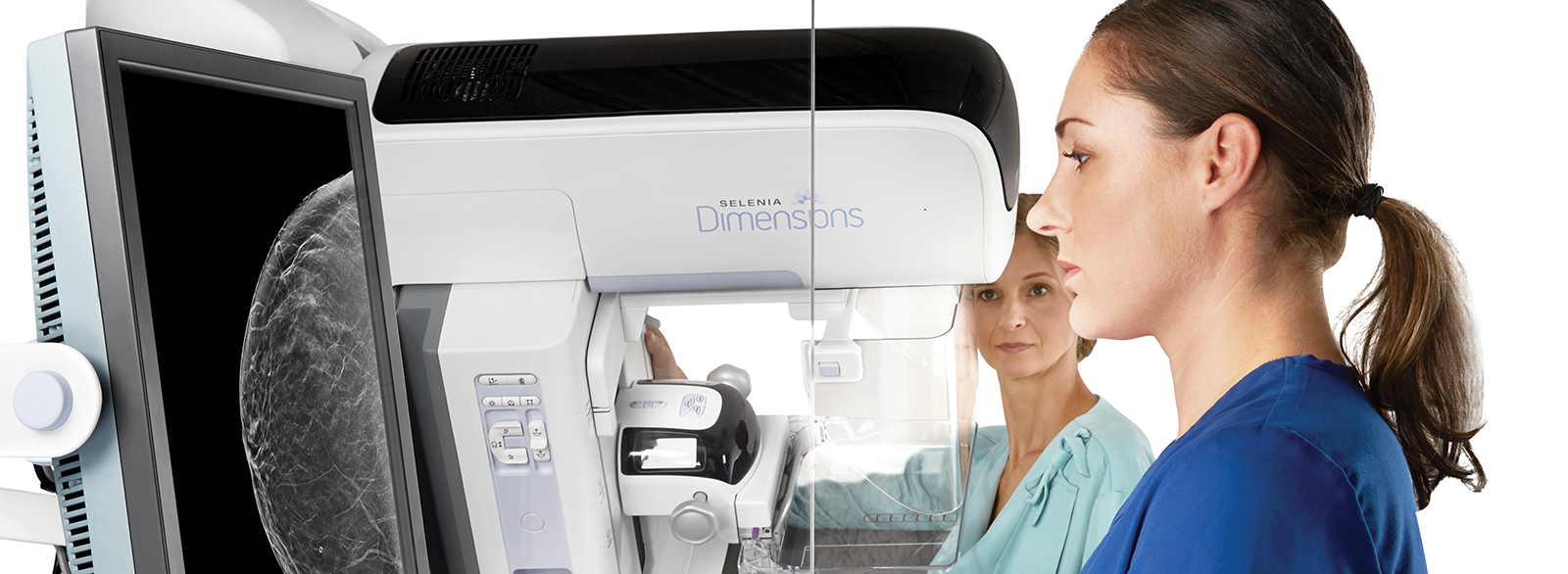
A 3D mammogram exam is very similar to a traditional mammogram. Just as with a digital mammogram, the technologist will position you, compress your breast under a paddle and take images from different angles. An exam may be used as a screening tool in conjunction with a traditional digital mammogram or may be used by itself for a diagnostic mammogram.

During the exam, the X-ray arm sweeps in a slight arc over the breast, taking multiple breast images in just seconds. Your doctor is then able to view your breast tissue in one millimeter layers. Instead of viewing all the complexities of your breast tissue in one flat image, the doctor can examine the tissue one page or “slice” at a time.

There is no additional compression required with the exam, and it only takes a few seconds longer for each view. The technologist will view the images at a computer workstation to ensure adequate images have been captured for review by a radiologist, who studies them and reports results to either your physician or directly to you.

Early detection is the key

Because our primary goal has always been to deliver the highest quality care to our patients, we have added digital breast tomosynthesis to our breast health services. We have chosen to offer the Genius 3D Mammography exam from Hologic because clinical studies show that it is a more accurate mammogram.

Please call The Breast Health Center at 812-669-1607 to schedule your exam.

Return to the Breast Health Center


Video Transcript

Ingrid Ott, MD: Here is a patient who's about 58 years old who came in for routine screening mammogram. She had no symptoms, no lump, no discharge, nothing, no family history.

I'll show you her 2D view first. This is a 2D view of her left breast. I don't really see anything here that stands out. There's dense tissue here, here, here, here, here. Nothing that really stands out as looking unusual compared to everything else.

Let's now look at her 3D image. I'm going to start at her head and move down to her feet. We're getting down towards the level of the nipple, and all of a sudden, we run into a 3-centimeter mass that was not apparent on the 2D image.

Well, she got lucky. That mass turned out to be a cyst, but while we were looking at that mass, we happened to notice that behind the mass, there was a spiculated mass, measured about 8 millimeters in size, and that spiculated mass turned out to be a cancer. That is about as small as we can find cancer in a breast that looks like this.

She was able to have conservative treatment, and within a few months, the whole episode had passed for her.

Narrator: No woman is safe from it. No one can prevent it. Our only weapon is time. That's why science is always looking for ways to find breast cancer sooner.

One new method is tomosynthesis or 3D mammography. It images the breast tissue in very thin layers, so a radiologist can see details that used to be obscure.

The research predicted that tomosynthesis would make a difference, but how much difference?

Agnes Holland, MD: Over the last four months, we have seen multiple cancers that we could only see on tomosynthesis.

Sherry Gage: Look at that. You can't see it on 2D. There was a very, very small cancer that you clearly could see the stellate lesion.

Gary Rose, MD: There was this really small cancer, it turned out that you couldn't see on the regular mammogram, but you could see it on the 3D mammogram.

Renate Tewaag, MD: We said, "Wow, there's something wrong," and we got some structures we had never seen in the 2D.

Agnes Holland, MD: All of a sudden, you see it, and you're like, "Oh, my gosh," and you look at the 2D, you're like, "Where is it?"

Julianne Greenberg, MD: It takes your breath away a little bit. You go, "There's something I really needed to see, and here it is for me, so clearly visible."

Mark Klein, MD: In our practice, and I think nationwide, it's been shown to increase the cancer detection rate, and not just by a little, by about a third, and that's a huge number in terms of improving a diagnostic test.

Sherry Gage: What's exciting to us is we've found close to 25 cancers that would have actually walked out the door, not being diagnosed.

Gary Rose, MD: If you're that one patient, that's all you need. Just finding it once is enough to make me feel a technology is worth it.

Narrator: The consensus seems to be that 3D mammography really does help physicians detect breast cancer sooner, but it also seems to raise their confidence in what they see. Without the ambiguity of overlapping tissue, many doctors find it easier to say when there's no cancer in the picture, and that makes a difference, too.

Stefano Ciatto, MD: Well, let's say that tomosynthesis sees more, sees more details. This is the first methodology, which increases sensitivity and the specificity at the same time. With all the other methodologies, as soon as you raise the sensitivity level, you drop specificity or the reverse, and this is the first with better on both sides.

Bagyam Raghavan, MD: One of the important things is not just telling a patient she has cancer, but when you tell the patient that she doesn't have cancer, the onus is on the radiologist, and that level of confidence you have to have.

Mark Klein, MD: We may see a mammogram that looks normal, and so when you go to talk to the woman, she'll ask you, "Well, how does it look?" You'll say, "Well, I think it's fine. We can't see everything on a mammogram. The overlapping tissue obscures some of the detail, and so there could be something there we don't see, but I want to reassure you that everything is probably fine." Well, probably is not good enough in medicine and certainly not from a patient's point of view.

Andrea Reszt, MD: A direct, clear way for the patient is always better than, "Perhaps," and, "We don't know," and, "It could be." After I thought that something was not very clear in the 2D mammography and I could tell her just after tomosynthesis that everything's okay with the breast, or that we are now sure that we have to do a biopsy, for example.

Mark Klein, MD: What tomosynthesis does, it, in many cases, allows the radiologist to say, "You know, Mrs. Jones, you have dense tissue, but I was able to see through the different layers, and things really look good, and I'm highly confident that things are okay." That makes the patient feel much better, and, of course, it makes the radiologist feel much better, because, after all, we do want to get them all right.

Julianne Greenberg, MD: It allows me to, at the end of my little session reading that mammogram, to feel much more confident that there is no lesion to be worried about, there's no reason for the patient to be called back, there's nothing worrisome for me to have anxiety for myself over in reading that mammogram, that I can confidently say to them, "You are fine. We'll see you in one year."

Edward Lipsit, MD: It helps me in both ways. The mammogram, the conventional mammogram, the two-dimensional mammogram that we talk about, it's a good test, but it's an imperfect test, and anything that helps me in being more sensitive and more specific, that's a tremendous advantage. Our screening mammogram callback rate will decrease. In my personal experience, my own recall rate has gone down by 22%.

Gary Rose, MD: With 3D, I can see there's nothing there, if there's not a real mass, or I can see there really is a mass. My recall rate has gone down dramatically.

Peter Dunner: What we're seeing is a different type of recall, if we have to, which we may be already aware that something is suspicious, and then the recall is to prove it by usually ultrasound or MRI.

Julianne Greenberg, MD: When we have to say that to a patient, and nobody really wants to hear that, but when we have to say, "You need a biopsy," we're more sure that there is a significant lesion there, and it has a high probability of representing a breast cancer.

Narrator: To some extent, radiologists still diverge over which women benefit most from 3D.

Radiologist: We first thought that doing tomosynthesis on just dense breast tissue patients was the way to go, but, actually, you can find cancers in fatty breasts, as well, on tomosynthesis that you can't see on 2D.

Aron Belfer, MD: For this lady, for example, that has a completely fatty breast, was a big difference. We can see there's a very small lesion here, about five millimeter in size, that appears early on the CC tomo view and on nothing else. This was invasive lobular carcinoma.

Stefano Ciatto, MD: The benefit is in both, and the drop in recall rate is very similar in dense and non-dense breasts, and the extra detection rate of cancer is slightly higher in dense breasts, so the benefit is bigger, but there is also benefit in non-dense breasts.

Prof. Rüdiger Schulz-Wendtland: When we see, for example, a lesion in a fatty breast, it's the same. It's important, too, to make an assessment for this. Therefore, I think, of course, the dense breast patients that is more important, but I think tomosynthesis will be for all patients.

Sherry Gage: We had two women that came in the door that said, "No, no. I have family history. I've been called back," but their breasts were fatty. We did the tomosynthesis exam, and in both of those women, we found one had a cancer and one had a very large radial scar. In both of these women, the 2D mammogram was 100% clear. We quickly changed our plan, and we offer tomosynthesis to every woman.

Radiologist: I think we're doing a disservice by not doing tomosynthesis on every patient.

Agnes Holland, MD: In our practice, we have a lot of female physicians, and amongst us, we have very varying breast densities ourselves, and we all get tomosynthesis, regardless of our breast density.

Julianne Greenberg, MD: Because the two major risk factors for developing breast cancer are being female and getting older, and 70% of breast cancers are diagnosed in women with no significant risk factor.

Narrator: 3D is happening. It's causing a shift in the way we think about breast cancer detection.

Ingrid Ott, MD: I do want you to know that this is not experimental. We have been waiting and watching this technology for a long time. Also, what this has done is decreased healthcare costs because, by the time this mass became apparent on a 2D view, I mean the spiculated mass, this would probably be maybe five or six years from now, by the time we could actually see it in that dense tissue, by then, she may well have had lymph node involvement in her axilla, she may have required a mastectomy.

The whole cost to healthcare in society would be much greater. Obviously, the women benefit, but we all benefit because it's a smaller cancer requiring less treatment and less long-term side effects and less healthcare costs. Don't forget, one in eight women will get breast cancer. We want to find them when they're eight millimeters.

Carole Rubin, MD: We don't prevent breast cancers. Our job is to detect it as early as possible, and 3D tomosynthesis is allowing us to do that. I think it will be the standard of care very soon.

41%


increase in invasive cancer detection

2D vs 3D Exams

2d vs 3d mammogram comparison

(Click to enlarge)

In a conventional 2D mammogram, if there appears to be an area of concern, the doctor may want to further investigate with another mammogram or a biopsy. Looking at the same breast tissue in slices from the 3D mammogram exam, the doctor can now determine if the tissue is in fact normal breast tissue creating the illusion of an abnormal area. In situations like these patients likely avoid a callback for an additional mammogram.

40%


reduction in false positives

Tomosynthesis Animation

Tomosynthesis scan thumbnail.
This brief animation (no sound) visually illustrates how the 3D Mammogram exam works.

Watch Video